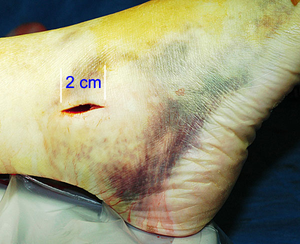
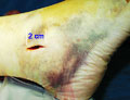
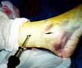
| The patient has had general anaesthesia, and the right ankle is prepped for surgery. A two centimeter incision is made over the end of the fibula. The extensive bruising around the ankle is normal in ankle fractures. |
 |
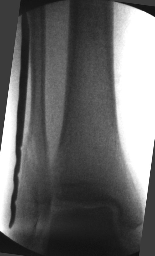
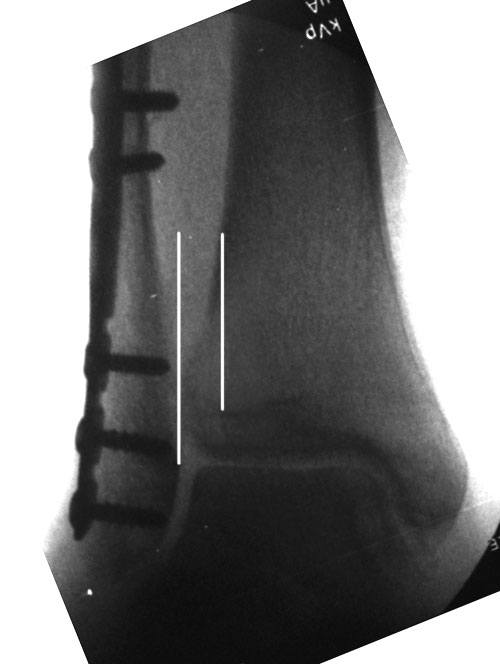
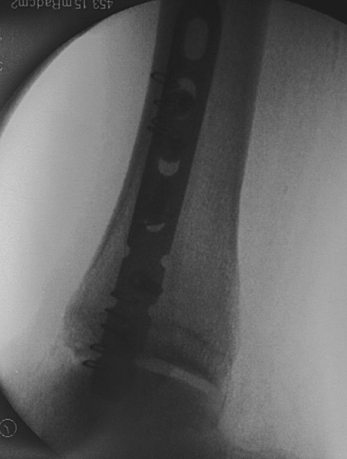
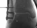
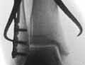
| The tissues next to the fibula are elevated and a titanium plate is slid in next to the bone. The xray confirms the correct location in the frontal plane. The fracture in the end of the fibula is clearly seen and is still displaced. |
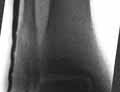 |
| The position of the plate is confirmed in the lateral plane. The plate is entirely inside the skin and cannot be seen without xray. |
 |
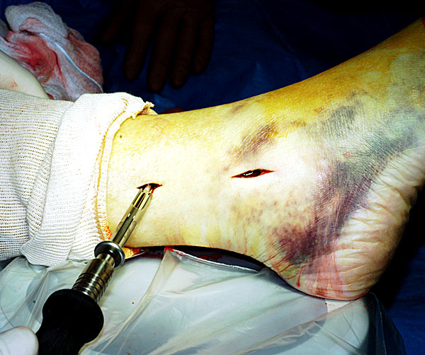
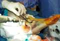
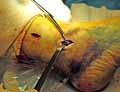
| A one centimeter incision over the top end of the plate is made and a single screw is placed percutaneously with the screwdriver. Special instruments protect the nearby tissues from injury. |
 |
| The fibula is reduced by the pressure of the plate and by positioning of the foot so that the ankle joint is perfectly aligned. The fracture is now barely visible. |
 |
| The screws in the bottom part of the plate are placed throught the lower incision. Each is verified with xray. |
 |
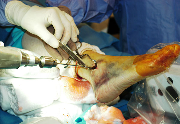
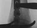
| The plate is in place. The space between the fibula and the tibia is abnormally increased due to damage to the syndesmosis ligament. The ankle is unstable. |
 |
| A caliper is placed on the fibula against the head of one of the screws to avoid damage to the bone. The syndesmosis is compressed into normal position. |
 |
| The space between the fibula and the tibia has been decreased and the bones are now in the correct position. A special screw will be inserted to hold it until it heals. |
 |
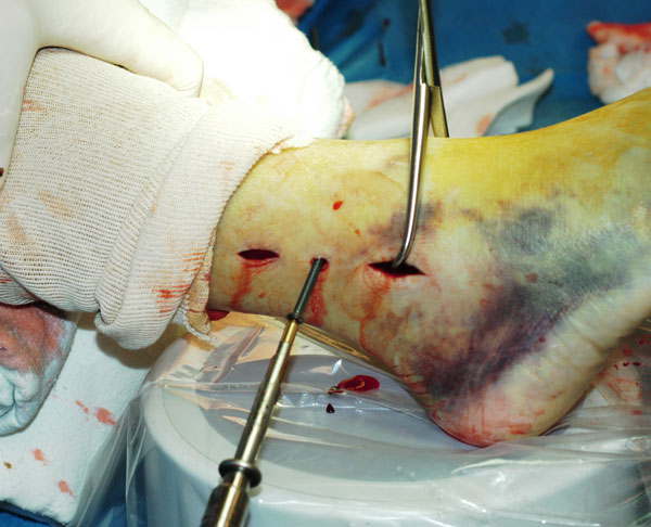
| A 5 millimeter incision is needed for the syndesmosis screw, midway between the top and bottom incisions. |
 |
| The position of the guide for the drill is verified by xray, since the entire plate under the skin. |
 |
| The drill hole for the screw is made and the correct length is chosen. |
 |
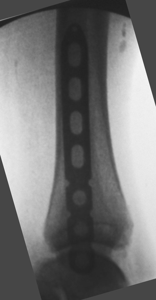
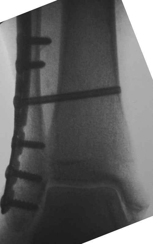
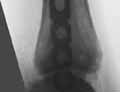
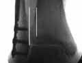
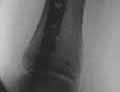
| This is the final xray appearance of the ankle after the repair. The fracture is perfectly aligned in the frontal plane and the syndesmosis between the bones is repaired. |
 |
| The final xray in the lateral plane. The plate is centered on the fibula and the joint is in perfect position. |
 |
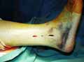
| The final appearance of the leg before the incisions are closed. The combined length of the incisions is 35 millimeters, about one quarter the length of a standard incision. |
 |
If you would like more information on how these treatments may help you with your particular health concerns, please